
Livestock Diseases
Influence of mastites on the physic-chemical features of goats' milk
- Abstracts: English Portuguese
Abstract in English:
Mille samples from tbree goat breads (Dusk Alpine, Saanem and Toggenburg) with milk production averaging 2.66 kg/day and from crossbred goats were examined during 12 months for physic-chemical features. The results for individual milk and the mixture were respectively: density 1,030.5 and 1,033.5 at 15ºC; acidity 15.67 and 15,84°D; average percentage of fat 4.00 and 4.1 %; total dry extract 12.42 and 12.95%; fatty dry extract 8.83 and 8.73%; cryoscopic indexes -0.569 and -0.585ºC. The average percentage of chlorides was normal (0.21 and 0.19%); lactose 3.27 and 3.42%; caseíne (PTN) 2.68 and 2.52%; total proteine (PTN) 3.59 and 3.43%. The results obtained with milk from goats with mastitis showed the following alterations caused by experimental infection by Staphylococcus aureus: lowered values for density (1,022.5 and 1,023.0 at 15ºC) and acidity (14.50 and 6.1 ºD); increased values for chlorides (0.34 and 0.39%); and reduced values for lactose (1.00 and 1.12%).
Abstract in Portuguese:
Amostras de leite de cabras de três raças selecionadas (Parda Alpina, Saanem e Toggenburg) com produtividade média de 2,66 kg/ dia e amostras de leite de cabras mestiças, foram examinados durante 12 meses, quanto às características físico-químicas, obtendo-se limites para o leite individual e de mistura, respectivamente, relativos à densidade entre 1.030,5 a 1.033,5 a 15ºC; a acidez entre 15,67 e 15,84°D; aos percentuais médios de gordura 4,00 e 4,1 %; ao extrato seco total entre 12,42 e 12,95%; ao extrato seco desengordurado entre 8,83 e 8,73%; aos índices criosc6picos entre -0,569 e -0,585ºC. As médias anuais referentes a porcentagem de cloretos se apresentaram dentro de limites discretamente acima da normalidade (0,21 e 0,19%); a lactose entre 3,27 e 3,42%; a caseína (PTN) entre 2,68 e 2,52% e o PTN total entre 3,33 e 3,43%; O leite proveniente de cabras com mastite, provocada experimentalmente com Staphylococcus aureus, apresentou alterações quanto à densidade (1.022,5 a 1.023,0 a 15ºC) e acidez (6,1º a 14,50°D); cloretos bastante aumentados (0,39 e 0,34%), e a lactose com valores bastante reduzidos (1,00 e 1,12%).
Experimental poisoning by Baccharis megapotamica var. megapotamica and var. weirii (Compositae) of rabbits
- Abstracts: English Portuguese
Abstract in English:
The dried and powdered aerial parts of Baccharis megapotamica Sprengel var. megapotamica and var. weirii (Baker) Barroso, plants which occur mainly in Southern Brazil, were toxic when administered by stomach tube to 68 rabbits; 42 of these died. The two varieties of the plant showed similar toxic properties, but there were a few differences. In both varieties the toxic symptoms were practically the sarne independently of its growing stage, the sex of the plants or its origin. However there were large differences in the lethal dose of the two varieties. It was between 2 and 4 g/kg for variety megapotamica and between 0,34 and 0,68 g/kg for var. weirii (dried plant material). Thus var. weirii was about 5 times more toxic than var. megapotamica. Fatal poisoning was acute with both varieties. In most cases symptoms caused by var. megapotamica were seen during 20 hours to 4 days; with var. weirii they were seen in most cases during less than 15 hours. Death occurred in most cases between 22 and 73 hours, and between 12 and 56 hours respectively after administration of the plant. The main symptoms shown by the rabbits intoxicated by var. megapotamica were anorexia and modified feces, mainly in the form of diarrhea, in most cases with the presence of mucus and blood. Anorexia was observed also in poisoning by var. weirii, but there were few cases of diarrhea and the faeces never contained mucus and blood. The main post-mortem findings in the rabbits poisoned by the two varieties were seen in the digestive tract, mainly in the stomach and caecum. The mucosa of the stomach was reddened, especially in the cranial part. The content of the caecum had a liquid-pasty consistency. Dark red slightly elevated and rough áreas were seen on the caecal mucosa. The caecal wall was frequently edematous. There were also lesions in the small and large intestine. In the small intestine lesions were seen only in poisoning by var. weirii and consisted of a reddened mucosa; in the colon and rectum severe lesions were seen only in poisoning by var. megapotamica and consisted of diphteroid necrosis. Both varieties produced livers that were almost always clearer than normal and slightly mottled. The histological lesions were necrosis and hemorrhages in the upper portion of the stomach mucosa, but necrosis of the parietal cells were more severe with var. megapotamica, being only slight with var. weirii. The mucosa of the small intestine only showed congestion and hemorrhages with var. weirii. The most important lesions were seen in the caecum, where there were hemorrhages and necrosis of the mucosa, and edema of the submucosa. Granulocytes were present, in most cases in small arnounts, but were generally more numerous with var. megapotamica, were some showed karyorrhexis; karyorrhexis was not seen after poisoning by var. weirii. In the initial part of the colon there were more hemorrhages and necrosis of the mucosa with var. weirii. However in the mucosa of the distal part of the colon and of the rectum, hemorrhages and necrosis with the presence of nuclear detritos (debris) on the surface of the mucosa, and necrosis with cariorrexia of the epithelial cells of the glands, were more common with var. megapotamica. Basically the sarne lesions were seen in the liver following poisoning by both varieties; there was severe tumefaction of the hepatic cells of the whole lobule, being especially severe in the periportal area. In the lymphatic tissue of the spleen and vermiform appendice of the caecum, necrosis characterized by cariorrexia of the lymphoid cells in the follicles, was produced by both varieties, but was more frequent and more pronounced with var. weirii. The two varieties, dried and milled, lost some of their toxicity, when kept in closed bottles; but when stored unmilled in cotton sacs, the plants kept their toxicity entirely. Rabbits, to which the two varieties were given, various times in sublethal doses with monthly intervals, did not adquire tolerance against the toxic properties of the plant.
Abstract in Portuguese:
As partes aéreas dessecadas e pulverizadas de Baccharis megapotamica Sprengel variedades megapotamica e weirii, plantas brasileiras conhecidas por conterem tricotecenos, foram administradas por via intragástrica a 68 coelhos, dos quais 42 morreram. O quadro clínico-patológico observado na intoxicação por ambas as variedades foi semelhante, porém a dose letal da var. megapotamica oscilou entre 2 e 4 g/kg, enquanto que a da var. weirii situou-se entre 0,34 e 0,68 g/kg, independentemente da fase de crescimento, do sexo da planta ou da procedência. Da mesma forma, a evolução nos casos fatais da intoxicação pela var. megapotamica foi mais longa (na maioria dos casos entre 7 e 32 horas) que a da var. weirii (geralmente menos de 15 horas). Na intoxicação pela var. megapotamica os principais sintomas foram anorexia e fezes alteradas, sobretudo diarréia, geralmente com presença de muco e sangue, já na intoxicação pela var. weirii havia também anorexia, porém a diarréia foi pouco freqüente e sem presença de muco ou sangue. Os achados de necropsia na intoxicação por ambas as variedades consistiram em avermelhamento da mucosa do estômago; ceco com áreas elevadas e rugosas, de coloração vermelho-escura, edema de parede e conteúdo líquido-pastoso; fígado mais claro e levemente mosqueado. Na intoxicação pela var. megapotamica havia ainda necrose difteróide no cólon e reto, enquanto que com a var. weirii observou-se avermelhamento da mucosa do intestino delgado. Histologicamente, havia, na intoxicação por ambas as variedades, necrose e hemorragias na parte superior da mucosa do estômago, porém necrose das células parietais foi bem mais acentuada na intoxicação pela var. megapotamica; no ceco, observaram-se hemorragias e necrose na mucosa, edema na submucosa e presença de polimorfonucleares que, na intoxicação pela var. megapotamica, em parte estavam em cariorrexia; no cólon, havia necrose e hemorragias, que na intoxicação pela var. weirii eram mais acentuadas na parte proximal, na intoxicação pela var. megapotamica eram mais acentuadas na parte distal e também presentes no reto; fígado com acentuada tumefação celular; no tecido linfóide (baço, apêndice vermiforme do ceco), necrose com cariorrexia, mais frequente e mais intensa na intoxicação pela var. weirii. Por 9 a 12 meses a planta dessecada inteira e em sacos de pano conservou integralmente a sua toxidez, porém quando guardada moída e em vidros de tampa plástica, perdeu um pouco de sua toxidez. Coelhos que receberam repetidamente doses subletais da planta, com intervalos mensais, não adquiriram tolerância à ação tóxica da planta.
Colony types and biochemical characteristics of Clostridium chauvoei isolated from cattle in Brazi
- Abstracts: English Portuguese
Abstract in English:
The morphological variation of colonies and biochemical characteristics of Clostridium chauvoei, isolated from the metatarsial and metacarpial bones of bovines with a history of blackleg, were investigated. The 59 cultures isolated contained 7 distinct morphological types of C. chauvoei and only 10 (16.95%) showed Type IV growth according to Zeissler (1928). All cultures hydrolized gelatin, had no proteolytic activíty and produced neither lecithinase nor lipase. Only 51 (86.44%) of the 59 cultures showed saccharolytic activity (lactose fermented). The biochemical profile was increased to 29 biochemical tests. The results indicate the morphological heterogenicity and biochemical variations of C. chauvoei in our environment.
Abstract in Portuguese:
A variação morfológica de colônias e do comportamento bioquímico de culturas de Clostridium chauvoei, isoladas de ossos metatarsianos e metacarpianos de bovinos com histórico clínico de carbúnculo sintomático, foi analisada. Das 59 culturas estudadas, observaram-se 7 tipos morfológicos distintos de C. chauvoei e somente 10 (16,95%) apresentaram crescimento do tipo IV. Todas as culturas isoladas hidrolizaram a gelatina, não apresentaram atividade proteolítica e nem produziram lecitinase e lipase. Das 59 culturas testadas 51 (86,44%) fermentaram a lactose (atividade sacarolítica). O perfil bioquímico foi ampliado para 29 provas bioquímicas. Estes resultados demostraram a heterogenicidade morfológica das colônias de C. chauvoei no nosso meio, apresentando variações nos seus parâmetros bioquímicos.
Histochemical diagnosis of actinomycotic-like granulomas in cattle from southern Brazil
- Abstracts: English Portuguese
Abstract in English:
The etiology of actinomycotic-like lesions from 254 lymph nodes of cattle from southern Brazil was determined by histochemistry. The diagnosis of these lesions in hematoxylin-eosin (H-E) preparations was based upon the pyogranulomatous inflammatory reaction with centrally located sulphur grains. The lesions stained by the MacCallum-Goodpasture technique showed 60.36% Gram-positive coccoid bacteria (Staphylococcus), 36.62% Gram-negative cocco-bacillus (Actinobacillus lignieresz) and 2.72% Gram-positive filamentous branching elements (Actinomyces bovis). Fite-Faraco stain demonstrated the acid-fast properties and Gomori stain, the morphological features to distinguish between Actinomyces bovis .and Nocardia sp. Gram-positive cocei were diagnosed as botryomycosis which occurred predominantly in prescapular, parotid and femoral lymph nodes. The presence of Gram-negative coccobacillary elements indicated Actinóbacillus sp., whereas the finding of Gram-positive filamentous branching elements indicated Actinomyces sp. These latter two were more frequently found in retropharyngeal and sublingual lymph nodes. Small differences in the morphology of the bacterial colonies may help to establish the etiological diagnosis in H-E preparations. The Gram method, however, allows an accurate distinction between Actinobacillus lignieresi and Actinomyces bovis in cases where fresh material for bacteriological isolation is no longer available. Of 467 other cases originally diagnosed as tuberculosis in gross inspection at the slaughter house, 10.23% were histologically confirmed as actinomycotic-like granulomas. This certainly causes economic losses due to the condemnation of carcasses.
Abstract in Portuguese:
Foi determinada a etiologia dos "granulomas actinomicóides" através da histoquímica em 254 materiais de linfonodos de bovinos do Rio Grande do Sul. O diagnóstico de granuloma actinomicóide foi baseado na inflamação piogranulomatosa, contendo, na porção central, os grânulos de enxofre, observados na coloração pela hematoxilina e eosina (H-E). Ao material com essa lesão foi aplicada a técnica de MacCallum-Ooodpasture que evidenciou uma incidência de 60,36% de bactérias cocoides Gram-positivas (Staphylococcus), 36,62% de coco-bacilos Gram-negativos (Actinobacillus lignieresz) e de 2,72% de elementos filamentosos, ramificados, Gram-positivos (Actinomyces bovis) como constituintes do grão de enxofre. Nos casos de actinomicose aplicaram-se as técnicas de Fite-Faraco e Prata de Gomori para a diferenciação entre Actinomyces bovis e Nocardia sp. Os casos de botriomicose foram vistos principalmente nos linfonodos pré-escapulares, parotídeos e femurais; os de actinobacilose e actinomicose nos retrofaringes e sublinguais. Pequenas diferenças do aspecto das colônias podem auxiliar no diagnóstico etiológico nas preparações coradas pela H-E, mas o emprego das técnicas de Gram permite a diferenciação entre Actinobacillus lignieresi e Actinomyces bovis quando não se dispõe de culturas. Adicionalmente observou-se pelo estudo histológico de outros 467 casos, que 10,23% deles foram diagnosticados como granuloma actinomicóide, porém interpretados macroscopicamente, na inspeção, como tuberculose o que certamente leva a prejuízos econômicos pela condenação das carcaças.
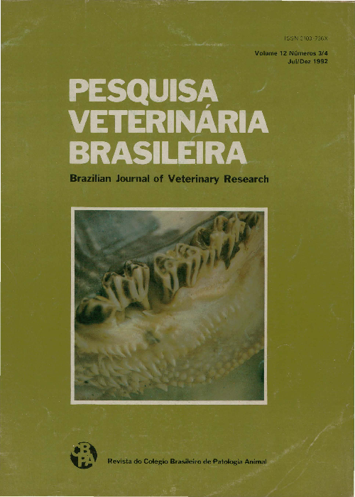
Efficiency of Virginiamycin for the recovery of calves from the periodontal disease "Cara inchada"
- Abstracts: English Portuguese
Abstract in English:
"Cara inchada" of young bovines (CI) is an important economic and health problem for cattle raising in certain areas of newly cultivated pastures in Brazil. Affected calves develop a purulent periodontitis that leads to loss of premolar teeth, mainly of the upper jaw, to emaciation and frequently to death. To study the efficiency of Virginiamycin for the recovery of calves kept on a farm under conditions which led to an prevalence of 61.5% of CI, an experiment was performed. Seventy seven calves with progressive periodontal lesions received orally during 8 consecutive weeks 0.032g of Virginiamycin/animal 3 times a week. Two control groups of diseased non-treated calves were used: the first consisting of 10 calves maintained within the treated group of CI animals and a second group of 95 calves affected by the disease. After the 8 week period the treated calves showed a very good recovery, but there was aggravation of symptoms in the 2 groups of nontreated animals, such as bad odor from the buccal cavity, weight loss, diarrhea and shedding of premolar teeth. It was concluded that Virginiamycin was efficient for the treatment of calves with CI.
Abstract in Portuguese:
A "cara inchada" dos bovinos (CI) constitui-se num importante problema econômico-sanitário da pecuária em determinadas áreas de pastagens recém-formadas no Brasil. Bezerros acometidos desenvolvem uma periodontite purulenta progressiva, levando à perda de dentes pré-molares maxilares. Quando mantidos em propriedades com alta incidência da doença, um considerável número dos animais afetados vem a morrer por emaciação. O presente experimento teve por finalidade verificar a eficácia da Virginiamicina na recuperação de bezerros com CI mantidos sob as condições que levaram a 61,5% de incidência da doença. Assim, 77 bezerros com lesões peridentárias progressivas receberam durante 8 semanas consecutivas, por via oral, 0,032g de Virginiamicina por dose, 3 vezes por semana. Como controle foram utilizados dois grupos: um constituído por 10 bezerros que permaneceram no mesmo lote de tratamento, porém sem receber a Virginiamicina, e outro, por 95 bezerros, todos mantidos nas mesmas condições sob as quais ocorreu a doença na propriedade. Ao final do período de administração do antibiótico, o lote tratamento apresentou melhora acentuada, ao passo que os animais dos outros dois lotes tiveram agravamento do quadro, com perda de dentes, mau odor da cavidade bucal, diarréia e emagrecimento progressivo. Concluiu-se que a Virginiamicina foi eficiente na recuperação de bezerros acometidos pela CI, quando estes parâmetros foram considerados.
Abortion caused by Ateleia glazioviana (Leg. Papilionoideae) in rats
- Abstracts: English Portuguese
Abstract in English:
Ateleia glazioviana Baillon (Leg. Papilionoideae), populary known as "timbó de Palmeira", is a tree found throughout Rio Grande do Sul, Brazil. It is reputed to induce abortion in cattle, to be toxic to fish and repellent to houshold insects. Evaluation of the effects of the hydroethanolic leaf extracts administered to pregnant female rats showed that the dichloromethane and amino acids extracts in doses of 100 mg.kg-1 caused a significant decrease in the number of progeny and a reduction in weight gains of pregnant females when compared with undosed controls. It is concluded that the dichloromethane extract, which contains apoiar substances (e.g. rutin and afrormosin) and the amino acids extract (proteinogenic and non proteinogenic amino acids) are responsible for the abortions or reabsorptions of foetusses in female rats.
Abstract in Portuguese:
Ateleia glazioviana Baillon, árvore pertencente à família Leguminosae Papilionoideae de ocorrência no Estado do Rio Grande do Sul, é conhecida popularmente como timbó de Palmeira, sendo considerada abortiva e tóxica para o gado, ictiotóxica e repelente de insetos. Neste estudo foram constituídos grupos experimentais com ratas grávidas que receberam diariamente, via intraperitonial, frações do extrato hidroetanólico de folhas de A. glazioviana na dose de 100 mg.kg-1. Foi observada redução significativa no número de filhotes nos grupos que receberam as frações diclorometano e aminoácidos. Redução significativa do ganho de peso das fêmeas gestantes foi verificada para os grupos tratados com as frações diclorometano, acetato de etila, n-butanol e aminoácidos. Conclui-se que as frações diclorometano, que contêm substâncias menos polares, tais como rotina e afrormosina, e aminoacidos, que contêm aminoácidos protéicos e não protéicos, são capazes de produzir abortos ou reabsorções e reduzir o desenvolvimento ponderal de ratas gestantes.


